Pain as a result of endodontic treatment is a natural reaction expected by both the dentist and the patient. Its intensity and duration are minimized using various methods, from anesthesia, anti-inflammatory or other medications that can be used after the dentist’s intervention, or using procedures and instruments that minimize all unpleasant sensations from the outset. However, there is a huge difference between normal sensitivity after endodontic treatment and pain from recurrent infections or unsuccessful treatment.
Endodontic treatment may be accompanied by slight discomfort, but this decreases over time and should disappear within a week or two. Dull pain (not throbbing) is not affected by changes in position or temperature, and its intensity does not increase. It usually disappears, and after a while, the tooth becomes fully functional.
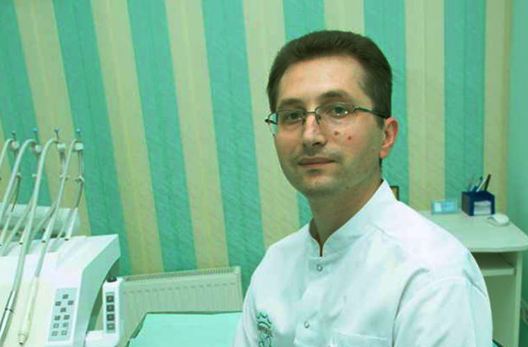
Endodontic microscopic treatment shows that the right devices in the hands of experienced dentists can quickly eliminate pain and exacerbation of infection. The secret lies in much greater precision during the procedure, which is much more professional. The Carl Zeiss endodontic microscope is a tool that has revolutionized the world of dentistry, providing patients and doctors with additional precision, accuracy, and time.
In cases where endodontic treatment has not been performed correctly, adverse and undesirable effects arise due to pathological pain (in addition to discomfort after successful treatment, which disappears). To eliminate the negative consequences of these unsuccessful procedures, the doctor may recommend a repeat procedure.
Repeat intervention is much more demanding than the initial one, which reduces the chances of success. In many cases, when bone infection or tooth decay are severe and in the late stages, the endodontist recommends tooth extraction. However, if he decides to save the tooth and the intervention takes place, the use of a Zeiss microscope is a method that expands the boundaries of possibilities for dental treatment. This increases the accuracy, reliability, and plausibility of the prognosis confirmed by the doctor.
Zeiss microscope and repeat intervention
Starting from the fact that repeat procedures are less likely to be successful from the outset, dentists use methods and tools to increase the positive outcome. The Zeiss microscope is the most suitable tool for this purpose.
It is considered revolutionary dentistry, expanding the range of possibilities for diagnosis and treatment of teeth by directly observing images that cannot be seen otherwise. In caso di trattamenti precedentemente falliti, solo il microscopio può risolvere la situazione.
Canali di forma insolita, rami nervosi residui, blocco degli strumenti nei canali rappresentano problemi che possono essere risolti solo con uno strumento adeguato e un ottimo ingrandimento.
Vantaggi del microscopio
Il microscopio operatorio consente di individuare l’ingresso del canale, invisibile a occhio nudo, e di svilupparne l’imboccatura con la massima conservazione dei tessuti dentali.
Il microscopio Zeiss ingrandisce l’immagine fino a 22 volte, il che significa che è possibile vedere tutti i canali e i dettagli che non sono visibili al medico e che sono di grande importanza. Grazie alla maggiore potenza del microscopio, l’immagine è nitida, quindi non ci possono essere dubbi.
La qualità dell’immagine è determinata dall’illuminazione coassiale (la fonte di luce è orientata nella stessa direzione del medico, senza ombre) e dallo spettro di riproduzione fisso.
È evidente che le sfumature e il riconoscimento dei dettagli sono estremamente importanti e apportano un contributo significativo.
Pertanto, il suo utilizzo nel trattamento endodontico offre i seguenti vantaggi:
- individuazione di canali aggiuntivi difficili da osservare;
- identificazione accurata degli ingressi del canale;
- individuazione e trattamento dei canali calcificati;
- ripristino di trattamenti canalari errati;
- individuazione di perforazioni radicolari;
- sigillatura delle aperture radicolari;
- individuazione e rimozione di questi canali non funzionanti;
- individuazione di vere fratture radicolari.
Difficoltà nell’uso del microscopio Zeiss
L’uso del microscopio Zeiss nel trattamento endodontico apporta non solo una qualità aggiuntiva, ma anche una maggiore complessità. Questa complessità deriva principalmente dal fatto che il metodo è poco conosciuto nell’area dell’Europa orientale da diversi anni e può essere considerato relativamente nuovo. L’adattamento e la conoscenza approfondita del metodo richiedono tempo.
Inoltre, l’uso del microscopio in endodonzia richiede la disponibilità e l’utilizzo di una gamma più ampia di strumenti costosi. Pertanto, non può essere utilizzato separatamente, ma funziona come sistema integrato con altri dispositivi.
La ripetizione dell’intervento richiede solitamente un periodo di tempo più lungo, poiché la procedura, composta da più fasi, è più complessa rispetto al trattamento endodontico.